The role of sacro-iliac joint magnetic resonance imaging in the diagnosis of axial spondyloarthritis: focus on differential diagnosis in women

Published: 11 September 2024
Abstract Views: 0
PDF: 0
Publisher's note
All claims expressed in this article are solely those of the authors and do not necessarily represent those of their affiliated organizations, or those of the publisher, the editors and the reviewers. Any product that may be evaluated in this article or claim that may be made by its manufacturer is not guaranteed or endorsed by the publisher.

Authors

Objective. To review the role of sacro-iliac magnetic resonance imaging (MRI) in the diagnosis of axial spondyloarthritis (AxSpA), with a focus on gender differences.

Methods. The experience of the authors and the results of an informal literature review are reported.

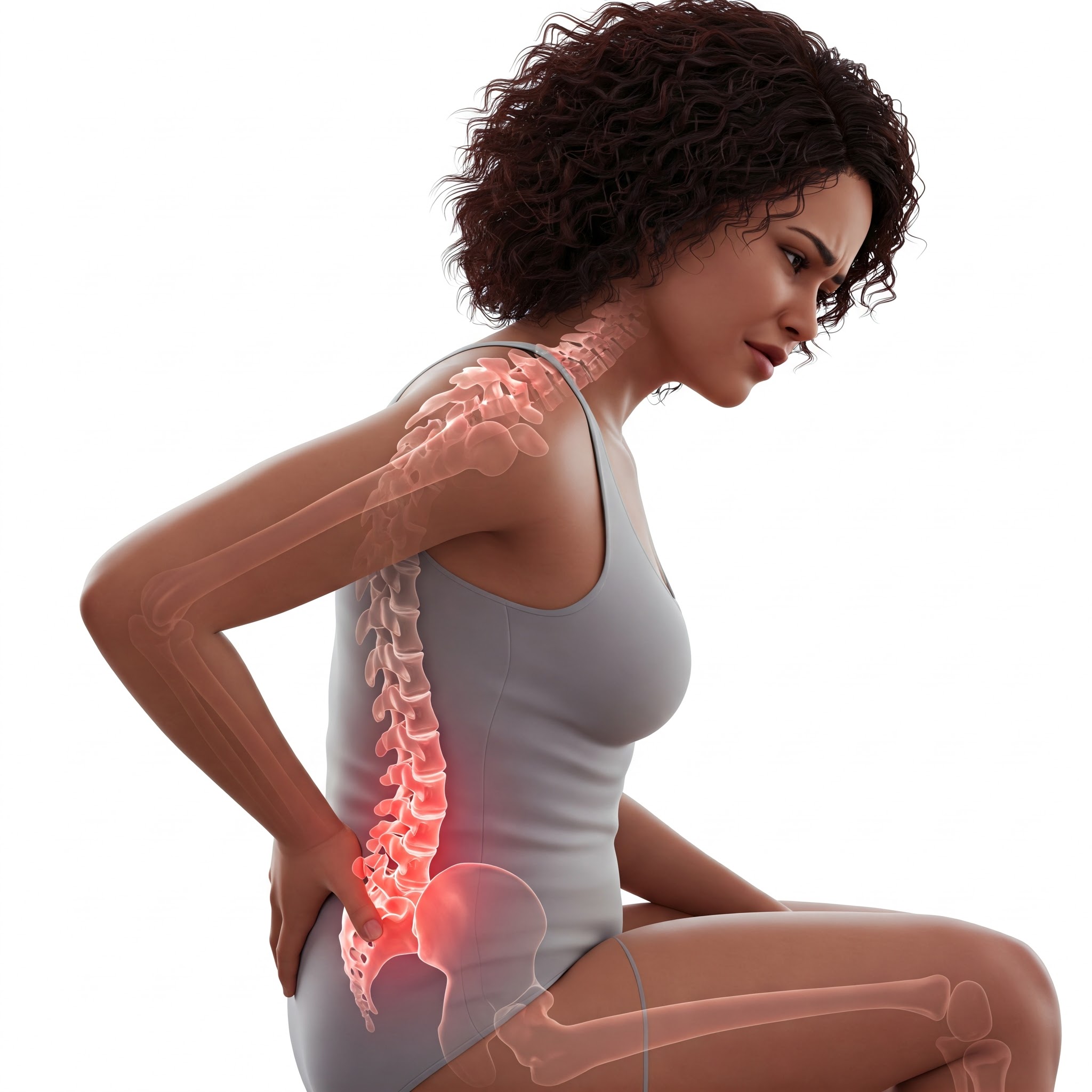
Results. Inflammatory changes of the sacro-iliac joint are the hallmark of AxSpA. Early, non-radiographic sacroiliitis may be diagnosed with MRI through the assessment of bone marrow edema (BMO) as well as concomitant structural damage. The MRI protocol should include three necessary sequences, i.e., fat-saturated T2-weighted sequences on two orthogonal planes, T1-weighted semi-coronal sequence, and fat-suppressed T1-weighted semi-coronal sequence. Inflammatory changes comprise required signs (BMO and/or osteitis) and additional signs, including synovitis (better defined as joint space enhancement), enthesitis, and capsulitis. Structural changes consist of erosions, sclerosis, fat metaplasia, and ankylosis. Due to mechanical axial strain, inflammatory changes in the sacro-iliac joint can be found in healthy individuals, runners, and patients with nonspecific low back pain. The prevalence of BMO is higher in women during pregnancy and postpartum, even 12 months after childbirth, but the extent and distribution of MRI findings may help in the differential diagnosis. Other challenges in the MRI diagnosis of sacroiliitis are subchondral T2 hyperintensity during developmental age, periarticular sclerosis in healthy subjects, or osteitis condensans ilii, and several pathological conditions that may mimic AxSpA, some of which are more frequently found in women.

Conclusions. The described diagnostic challenges impose a multidisciplinary approach combining imaging findings with clinical and laboratory data.

Dimensions

Altmetric

PlumX Metrics

Downloads

Download data is not yet available.

Citations

Navarro-Compán V, Sepriano A, El-Zorkany B, van der Heijde D. Axial spondyloarthritis. Ann Rheum Dis 2021; 80: 1511-21.
Rudwaleit M, Landewé R, van der Heijde D, Listing J, Brandt J, Braun J, et al. The development of Assessment of SpondyloArthritis International Society classification criteria for axial spondyloarthritis (part I): classification of paper patients by expert opinion including uncertainty appraisal. Ann Rheum Dis 2009; 68: 770-6.
Lambert RG, Bakker PA, van der Heijde D, Weber U, Rudwaleit M, Hermann KG, et al. Defining active sacroiliitis on MRI for classification of axial spondyloarthritis: update by the ASAS MRI working group. Ann Rheum Dis 2016; 75:1958-63.
de Hooge M, Diekhoff T, Poddubnyy D. Magnetic resonance imaging in spondyloarthritis: friend or foe?. Best Pract Res Clin Rheumatol 2023; 37:101874.
Rudwaleit M, Jurik AG, Hermann KG, Landewé R, van der Heijde D, Baraliakos X, et al. Defining active sacroiliitis on magnetic resonance imaging (MRI) for classification of axial spondyloarthritis: a consensual approach by the ASAS/OMERACT MRI group. Ann Rheum Dis 2009; 68: 1520-7.
Sudoł-Szopińska I, Jurik AG, Eshed I, Lennart J, Grainger A, Østergaard M, et al. Recommendations of the ESSR arthritis subcommittee for the use of magnetic resonance imaging in musculoskeletal rheumatic diseases. Semin Musculoskelet Radiol 2015; 19: 396-411.
de Hooge M, van den Berg R, Navarro-Compán V, van Gaalen F, van der Heijde D, Huizinga T, et al. Magnetic resonance imaging of the sacroiliac joints in the early detection of spondyloarthritis: no added value of gadolinium compared with short tau inversion recovery sequence. Rheumatology 2013; 52: 1220-4.
Herregods N, Jaremko JL, Baraliakos X, Dehoorne J, Leus A, Verstraete K, et al. Limited role of gadolinium to detect active sacroiliitis on MRI in juvenile spondyloarthritis. Skeletal Radiol 2015; 44: 1637-46.
Gentili F, Cantarini L, Fabbroni M, Nigri A, Mazzei FG, Frediani B, et al. Magnetic resonance imaging of the sacroiliac joints in SpA: with or without intravenous contrast media? A preliminary report. Radiol Med 2019; 124: 1142-50.
Maksymowych WP, Lambert RG, Østergaard M, Pedersen SJ, Machado PM, Weber U, et al. MRI lesions in the sacroiliac joints of patients with spondyloarthritis: an update of definitions and validation by the ASAS MRI working group. Ann Rheum Dis 2019; 78: 1550-8.
Diekhoff T, Poddubnyy D, Proft F, Ziegeler K, Deppe D, Niedermeier C, et al. New bone formation at the sacroiliac joint in axial spondyloarthritis: characterization of backfill in MRI and CT. Rheumatology 2023; 62: 3893-8.
Nygaard A, Jurik AG, Lund C, Schiøttz-Christensen B. The incidence of bone marrow oedema at the sacroiliac joints in a non-rheumatological population - a retrospective cohort study. BMC Musculoskelet Disord 2019; 20: 590.
Weber U, Jurik AG, Zejden A, Larsen E, Jørgensen SH, Rufibach K, et al. Frequency and anatomic distribution of magnetic resonance imaging features in the sacroiliac joints of young athletes: exploring “background noise” toward a data-driven definition of sacroiliitis in early spondyloarthritis. Arthritis Rheumatol 2018; 70: 736-45.
de Winter J, de Hooge M, van de Sande M, de Jong H, van Hoeven L, de Koning A, et al. Magnetic resonance imaging of the sacroiliac joints indicating sacroiliitis according to the Assessment of SpondyloArthritis International Society definition in healthy individuals, runners, and women with postpartum back pain. Arthritis Rheumatol 2018; 70: 1042-8.
Eshed I, Miloh-Raz H, Dulitzki M, Lidar Z, Aharoni D, Liberman B, et al. Peripartum changes of the sacroiliac joints on MRI: increasing mechanical load correlating with signs of edema and inflammation kindling spondyloarthropathy in the genetically prone. Clin Rheumatol 2015; 34: 1419-26.
Kiil RM, Weber U, Loft AG, Maimburg RD, Jurik AG. Evolution of magnetic resonance imaging lesions at the sacroiliac joints during and after pregnancy by serial magnetic resonance imaging from gestational week twenty to twelve months postpartum. Arthritis Rheumatol 2023; 75: 1166-75.
Renson T, Depicker A, De Craemer AS, Deroo L, Varkas G, de Hooge M, et al. High prevalence of spondyloarthritis-like MRI lesions in postpartum women: a prospective analysis in relation to maternal, child and birth characteristics. Ann Rheum Dis 2020; 79: 929-34.
Storey T, Borgia RE, Merritt P, Woolnough L. Normal magnetic resonance imaging appearance of marrow adjacent to the sacroiliac joints in children during development. J Comput Assist Tomogr 2022; 46: 91-6.
Borlandelli E, Ciaffi J, Festuccia G, Facchini G, Miceli M, Brusi V, et al. Osteitis condensans ilii: prevalence and characteristics of a neglected mimic of sacroiliitis. Clin Rheumatol 2022; 41: 483-90.
Mir R, Dragan AD, Mistry HB, Tsang YM, Padhani AR, Hoskin P. Sacral insufficiency fracture following pelvic radiotherapy in gynaecological malignancies: development of a predictive model. Clin Oncol (R Coll Radiol) 2021; 33: e101-9.
Wu YF, Lu K, Girgis C, Preda M, Preda V. Postpartum bilateral sacral stress fracture without osteoporosis-a case report and literature review. Osteoporos Int 2021; 32: 623-31.

How to Cite

Besutti, G., Marvisi, C., Muratore, F., & Spaggiari, L. (2024). The role of sacro-iliac joint magnetic resonance imaging in the diagnosis of axial spondyloarthritis: focus on differential diagnosis in women. Reumatismo, 76(3). https://doi.org/10.4081/reumatismo.2024.1768