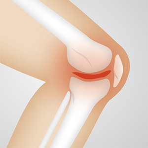
Magnetic resonance imaging as a structural refinement to the American College of Rheumathology clinical classification criteria for knee osteoarthritis

Published: 29 December 2022
Abstract Views: 970
PDF: 612
Publisher's note
All claims expressed in this article are solely those of the authors and do not necessarily represent those of their affiliated organizations, or those of the publisher, the editors and the reviewers. Any product that may be evaluated in this article or claim that may be made by its manufacturer is not guaranteed or endorsed by the publisher.
All claims expressed in this article are solely those of the authors and do not necessarily represent those of their affiliated organizations, or those of the publisher, the editors and the reviewers. Any product that may be evaluated in this article or claim that may be made by its manufacturer is not guaranteed or endorsed by the publisher.
Similar Articles
- I. Olivieri, E. Scarano, A. Padula, S. D'Angelo, IMAGING OF PSORIATIC ARTHRITIS , Reumatismo: Vol. 59 No. s1 (2007)
- M. Lorenzin, A. Ortolan, S. Vio, M. Favero, F. Oliviero, M. Zaninotto, C. Cosma, C. Lacognata, L. Punzi, R. Ramonda, Biomarkers, imaging and disease activity indices in patients with early axial spondyloarthritis: the Italian arm of the SpondyloArthritis-Caught-Early (SPACE) Study , Reumatismo: Vol. 69 No. 2 (2017)
- A. Bortoluzzi, M. Padovan, I. Farina, E. Galuppi, F. De Leonardis, M. Govoni, Therapeutic strategies in severe neuropsychiatric systemic lupus erythematosus: experience from a tertiary referral centre , Reumatismo: Vol. 64 No. 6 (2012)
- A. Marchesoni, F. Cantini, Classification and clinical assessment , Reumatismo: Vol. 64 No. 2 (2012)
- J.H. Villafañe, R. Cantero-Tellez, S. Negrini, P. Berjano, From art to science: the functional damage due to thumb osteoarthritis finely described by Velazquez 300 years before its clinical description , Reumatismo: Vol. 69 No. 3 (2017)
- G. Filippou, A. Adinolfi, A. Delle Sedie, E. Filippucci, A. Iagnocco, F. Porta, L. M. Sconfienza, S. Tormenta, V. Di Sabatino, V. Picerno, B. Frediani, Radiologists and rheumatologists on performing and reporting shoulder ultrasound: from disagreement to consensus , Reumatismo: Vol. 66 No. 3 (2014)
- F. Salaffi, A. Ciapetti, M. Carotti, The sources of pain in osteoarthritis: a pathophysiological review , Reumatismo: Vol. 66 No. 1 (2014)
- F. Paparo, E. Fabbro, G. Ferrero, R. Piccazzo, M. Revelli, D. Camellino, G. Garlaschi, M.A. Cimmino, Imaging studies of crystalline arthritides , Reumatismo: Vol. 63 No. 4 (2011): Special issue • Microcrystalline Arthritis
- C. Botsios, P. Sfriso, C. Grava, P. Ostuni, M. Andretta, A. Tregnaghi, P. Zucchetta, PF. Gambari, Imaging in major salivary gland diseases , Reumatismo: Vol. 53 No. 3 (2001)
- N. Romano, A. Fischetti, V. Prono, S. Migone, F. Barbieri, C. Pizzorni, G. Garlaschi, M.A. Cimmino, Plantar pain is not always fasciitis , Reumatismo: Vol. 69 No. 4 (2017)
<< < 1 2 3 4 5 6 7 8 9 10 > >>
You may also start an advanced similarity search for this article.

 https://doi.org/10.4081/reumatismo.2022.1534


