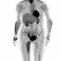
The role of PET in a clinically silent and ultrasound negative synovitis in the course of rheumatoid arthritis - a case report

Submitted: 23 September 2019
Accepted: 19 February 2021
Published: 19 April 2021
Accepted: 19 February 2021
Abstract Views: 3305
PDF: 445
Publisher's note
All claims expressed in this article are solely those of the authors and do not necessarily represent those of their affiliated organizations, or those of the publisher, the editors and the reviewers. Any product that may be evaluated in this article or claim that may be made by its manufacturer is not guaranteed or endorsed by the publisher.
All claims expressed in this article are solely those of the authors and do not necessarily represent those of their affiliated organizations, or those of the publisher, the editors and the reviewers. Any product that may be evaluated in this article or claim that may be made by its manufacturer is not guaranteed or endorsed by the publisher.
Similar Articles
- S. Canestri, M.C. Totaro, E. Serone, B. Tolusso, D. Frezza, E. Gremese, G. Ferraccioli, Association between the response to B cell depletion therapy and the allele*2 of the HS1,2A enhancer in seropositive rheumatoid arthritis patients , Reumatismo: Vol. 64 No. 6 (2012)
- S.L. Bello, L. Serafino, C. Bonali, N. Terlizzi, C. Fanizza, C. Anecchino, G. Lapaldula, Incidence of influenza-like illness into a cohort of patients affected by chronic inflammatory rheumatism and treated with biological agents , Reumatismo: Vol. 64 No. 5 (2012)
- C.-N. Lo, G. Xia, B.P. Leung, The effect of nerve mobilization exercise in patients with rheumatoid arthritis: a pilot study , Reumatismo: Vol. 69 No. 3 (2017)
- A. Marino, I. Pagnini, S. Savelli, D. Moretti, G. Simonini, R. Cimaz, Elbow monoarthritis: an atypical onset of juvenile idiopathic arthritis , Reumatismo: Vol. 64 No. 3 (2012)
- M. Catanoso, N. Pipitone, C. Salvarani, Epidemiology of psoriatic arthritis , Reumatismo: Vol. 64 No. 2 (2012)
- M. S. Dag, I. H. Turkbeyler, Z. A. Ozturk, B. Kısacık, E. Tutar, A. Kadayıfçı, Cytomegalovirus ileocolitis in a rheumatoid arthritis patient: case report and literature review , Reumatismo: Vol. 67 No. 1 (2015)
- M. Capraro, M. Dalla Valle, M. Podswiadek, P. De Sandre, E. Sgnaolin, R. Ferrari, The role of illness perception and emotions on quality of life in fibromyalgia compared with other chronic pain conditions , Reumatismo: Vol. 64 No. 3 (2012)
- Ş. Kobak, A. Berdeli, Fas/FasL promoter gene polymorphism in patients with rheumatoid arthritis , Reumatismo: Vol. 64 No. 6 (2012)
- F. Zare, M. Dehghan-Manshadi, A. Mirshafiey, The signal transducer and activator of transcription factors lodge in immunopathogenesis of rheumatoid arthritis , Reumatismo: Vol. 67 No. 4 (2015)
- M. Rossini, G. D'Avola, M. Muratore, N. Malavolta, F. Silveri, G. Bianchi, B. Frediani, G. Minisola, M. L. Sorgi, M. Varenna, R. Foti, G. Tartarelli, G. Orsolini, S. Adami, Regional differences of vitamin D deficiency in rheumatoid arthritis patients in Italy , Reumatismo: Vol. 65 No. 3 (2013)
You may also start an advanced similarity search for this article.

 https://doi.org/10.4081/reumatismo.2021.1253


