The role of PET in a clinically silent and ultrasound negative synovitis in the course of rheumatoid arthritis - a case report

Submitted: 23 September 2019
Accepted: 19 February 2021
Published: 19 April 2021
Abstract Views: 3100
PDF: 346
Publisher's note
All claims expressed in this article are solely those of the authors and do not necessarily represent those of their affiliated organizations, or those of the publisher, the editors and the reviewers. Any product that may be evaluated in this article or claim that may be made by its manufacturer is not guaranteed or endorsed by the publisher.

Authors

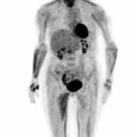
We present a case report of a rheumatoid arthritis patient, who underwent a PET scan, which revealed inflammation of multiple joints, which was missed by both physical and ultrasound examinations. A 55-year old woman with a long-term rheumatoid arthritis, who had undergone arthroplasty of the left knee in the past, consulted with the rheumatologist for pain in the left knee. The physical examination revealed signs of inflammation in the left knee and right elbow. The inflammatory parameters were high. Ultrasound showed intraarticular effusion without signs of active synovitis in the left knee. The ultrasound assessment of the other joints (hands, wrists and feet) was also negative for active synovitis, while positron-emission tomography (PET) revealed increased glucose metabolism at the level of the medial side of the left knee, left radio-ulno-carpal joint, I-II-III metacarpo-phalangeal joints bilaterally, right II metatarso-phalangeal joint, and left II-III metatarso-phalangeal joints. This case report demonstrates that PET might be more sensitive than ultrasound in detecting subclinical joint inflammation.

Dimensions

Altmetric

PlumX Metrics

Downloads

Download data is not yet available.

Citations

Colebatch AN, Edwards CJ, Østergaard M, et al. EULAR recommendations for the use of imaging of the joints in the clinical management of rheumatoid arthritis. Ann Rheum Dis. 2013; 72: 804-14. DOI: https://doi.org/10.1136/annrheumdis-2012-203158
Lee SJ, Jeong JH, Lee CH, et al. Development and validation of an 18 F-FDG PET/CT-based tool for the evaluation of joint counts and disease activity in patients with rheumatoid arthritis. Arthritis Rheumatol. 2019; 71: 1232-40. DOI: https://doi.org/10.1002/art.40860
Beckers C, Jeukens X, Ribbens C, et al. (18)F-FDG PET imaging of rheumatoid knee synovitis correlates with dynamic magnetic resonance and sonographic assessments as well as with the serum level of metalloproteinase-3. Eur J Nucl Med Mol Imaging. 2006; 33: 275-80. DOI: https://doi.org/10.1007/s00259-005-1952-3
dos Anjos DA, da Mota LM. Positron emission tomography with (18)F-FDG in the evaluation of patients with rheumatoid arthritis - a systematic review (in Portuguese). Rev Bras Reumatol. 2014; 54: 474-82. DOI: https://doi.org/10.1016/j.rbre.2014.07.003
Bhattarai A, Nakajima T, Sapkota S, et al. Diagnostic value of 18F-fluorodeoxyglucose uptake parameters to differentiate rheumatoid arthritis from other types of arthritis. Medicine (Baltimore) 2017; 96: e7130. DOI: https://doi.org/10.1097/MD.0000000000007130
Gent YY, Ter Wee MM, Voskuyl AE, et al. Subclinical synovitis detected by macrophage PET, but not MRI, is related to short-term flare of clinical disease activity in early RA patients: an exploratory study. Arthritis Res Ther. 2015; 17: 266. DOI: https://doi.org/10.1186/s13075-015-0770-7
Suto T, Okamura K, Yonemoto Y, et al. Prediction of large joint destruction in patients with rheumatoid arthritis using 18F-FDG PET/CT and disease activity score. Medicine (Baltimore) 2016;95:e2841. DOI: https://doi.org/10.1097/MD.0000000000002841

How to Cite

Skoczynska, M., Figus, F., Arena, V., Massazza, G., & Iagnocco, A. (2021). The role of PET in a clinically silent and ultrasound negative synovitis in the course of rheumatoid arthritis - a case report. Reumatismo, 73(1), 64–66. https://doi.org/10.4081/reumatismo.2021.1253